Pathologic features, nomenclature and diagnosis of small vessel vasculitis

Dr. Charles Jennette

May 14, 1997

Dr. Jennette
Charles Jennette, M.D.
Dr. Jennette is Professor of Pathology and Laboratory Medicine at the University of North Carolina at Chapel Hill.

Synchronized Real Media from Dr. Jennette's Presentation

28.8 modem
click here to listen!
This page is PART TWO. You can scroll through the talk while listening to the audio. There are about 30 slides to this talk in all, 15 in each part. Download this whole page before starting up the Real Player! The page will automatically scroll to the slide being discussed.


Part Two: Antineutrophil Cytoplasmic Antibodies Clarify the Situation

Dr. Jennette


Granular IgA staining in the walls of small dermal vessels in a patient with Henoch-Schönlein purpura.
The role that immune complexes play is appreciated
Fortunately some other modalities of diagnostic evaluation came into the picture in the 60s, in particular, and 70s. Immunohistology demonstrated that at least some of these patients with small-vessel vasculitis had immune complexes in the vessel walls, for example, here in the dermis of a patient with leukocytoclastic angiitis histologically and purpura clinically.


A number of immune complex vasculitides were subsequently recognized. Henoch-Schönlein purpura has been redefined and restricted to those patients with IgA-dominant immune complexes as the cause of their small-vessel vasculitis. By that definition, it occurs primarily in children and usually has a good outcome. Cryoglobulinemia vasculitis was recognized by McClusky and his associates initially and, of course, is the result of cryoglobulins, especially Types II and III cryoglobulins localizing in small vessels and inciting inflammation. Lupus can, on occasion, have a systemic vasculitis; rheumatoid arthritis patients occasionally have an immune complex vasculitis; serum sickness vasculitis; infection-associated vasculitis; paraneoplastic vasculitities; some drug-induced vasculitides can show evidence for immune complex deposition.


Patterns of glomerular immunostaining: LEFT: granular (immune complex); MIDDLE: linear (anti-GBM), and RIGHT: absent (pauci-immune).
Pauci-immune vasculitis is appreciated
For a while it was considered that possibly most, maybe even all, small-vessel vasculitides were caused by immune complex deposition or anti-basement membrane antibodies. And, of course, when you look at the glomerular involvement, which is frequent in patients with small-vessel vasculitis, you certainly do see some patients with granular staining indicative of an immune complex disease and linear staining indicative of anti-basement membrane disease. However, it became apparent to any number of investigators that the involved vessels other than glomeruli as well as the glomeruli in many patients with small-vessel vasculitis, especially patients with Wegener's granulomatosis and microscopic polyangiitis, did not have staining for immunoglobulin or complement. So this paucity of staining was somewhat of a problem in explaining the pathogenesis.


Prevalence of immune complexes in vasculitis
And in fact, these patients with pauci-immune vasculitis were quite common. For example, in this analysis of patients evaluated in my laboratory, you can see that the pauci-immune category is about equal to the immune complex category and much more frequent that anti-GBM disease if you look at all patients with inflammatory glomerulonephritis. And as you look at more and more severe glomerulonephritis, the frequency of this pauci-immune category goes up even more. So if you look at those patients who have greater than 50 percent of their glomeruli involved with crescents, this pauci-immune category is the most common. And, in fact, if you look at those few patients in whom you see necrotizing vasculitis in addition to glomerulonephritis in the kidney, there is a strong predilection here for the pauci-immune category.


Pauci-immune glomerulonephritis and ANCA positivity
Also in older patients, as has been seen here in Scandinavia by Pedersen and her associates in Stockholm, the frequency of this pauci-immune pattern and in fact this ANCA-associated disease pattern goes up. You can see here in this particular cohort, 90 percent of the patients were ANCA positive. So the pauci-immune category of glomerulonephritis and small-vessel vasculitis almost equates with ANCA vasculitis and glomerulonephritis.


As will be discussed later in detail by Jörgen Wieslander, ANCA specificity, as is well known to this group, in patients with glomerulonephritis vasculitis breaks down primarily into patients with anti-proteinase 3 specific antibodies that react with the cytoplasm and cause the C ANCA staining pattern and anti-myeloproxidase specific autoantibodies that react with antigen that is redistributed to the nucleus artifactually during preparation of the substrate if you use alcohol-fixed neutrophils. So these two ANCA specificities then mark a subset of small-vessel vasculitis that probably have a related pathogenesis.


Diagram depicting that some patients with ANCA-glomerulonephritis have no evidence for system vasculitis, however, most patients do.
ANCA vasculitis: glomerular and extrarenal involvement patterns
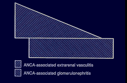
If you look at all patients with ANCA- positive glomerulonephritis, some of them apparently have involvement of no other tissues from all of the evidence that you can collect. But most patients with ANCA-positive glomerulonephritis have evidence for involvement of other viscera. The diagnostic challenge in these patients is to categorize those patients into some specific set if possible. This may be a somewhat academic exercise because, as we will discuss some later, the treatment may be very similar if not identical for all of these categories.


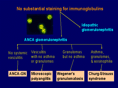
So the main step is initially realizing that it is an ANCA-positive necrotizing small-vessel vasculitis and possibly proceeding with treatment at that point and trying to categorize them further. If there is no systemic involvement, just ANCA glomerulonephritis, some prefer the term renal-limited vasculitis. If there is involvement of the respiratory tract or elsewhere with granulomatous inflammation, then Wegener's granulomatosis is the proper designation if there is no history of asthma. If there is a history of asthma, then Churg-Strauss syndrome. In these patients, the eosinophilia is usually more than 10 percent. If there is no evidence for Wegener's granulomatosis or Churg-Strauss syndrome, then the term that I would suggest is microscopic polyangiitis. I believe this is preferable to microscopic polyarteritis because many of these patients have no apparent involvement of arteries at all. They may have pulmonary capillaritis, they may have postcapillary venulitis in the skin, they may have glomerulonephritis, but they may not have arteritis. So the term microscopic polyangiitis is a better generic term.
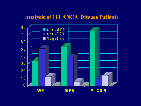

ANCA specificities
With respect to the association of the autoantibody specificities, various cohorts have been analyzed. This is an analysis of 111 patients from our cohort of ANCA-diseased patients. There is a predominance of the anti-PR3 specificity in patients with the Wegener's granulomatosis phenotype, but at least in our experience at this point, about one-third of these patients have anti-MPO specificity. In the microscopic polyangiitis patients, there is about an equal frequency of anti-MPO and anti-PR3 antibodies with possibly a little bit of predominance of anti-MPO specificity. In the patients with renal-limited disease, there is a very striking predilection for the anti-MPO.


Chapel Hill nomenclature system
I would suggest that the categorization of small-vessel vasculitis in patients with renal disease can usually be accommodated by what is sometimes referred to as the Chapel Hill nomenclature system, which categorizes some of these major forms of small-vessel vasculitis as Wegener's granulomatosis, Churg-Strauss syndrome, microscopic polyangiitis, all associated with ANCA; Henoch-Schönlein purpura, with IgA dominant immune complexes; cryoglobulinemic vasculitis, with circulating cryoglobulins and cryoglobulins in the tissues; and then occasional patients who have small-vessel vasculitis just confined to the skin. These patients may have a self-limited, benign course, but some of these patients will ultimately be found to have systemic involvement including the kidney.


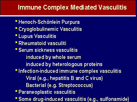
Vasculitides not on the above list
Of course, there are other forms of small-vessel vasculitis not accommodated in this particular list that I've described earlier: lupus- associated vasculitis, some forms of drug-induced vasculitis, infection-associated vasculitis. But in all of these circumstances, I think the initial approach should be to recognize that your patient has a small-vessel vasculitis and then as best you can to pursue additional diagnostic procedures, for example immunohistology on biopsy tissue, serology, to put them into a more specific category because that will impact substantially on the appropriate treatment regimen.

Thank you.

References

1. Godman G, Churg J. Wegener's granulomatosis. Pathology and review of the literature. Arch Pathol Lab Med 1954;58:533-553.

2. Jennette JC, Falk RJ, Andrassy K, et al. Nomenclature of systemic vasculitides: The proposal of an international consensus conference. Arthritis Rheum 1994;37:187-192.

3. Kallenberg CGM, Brouwer E, Weening JJ, Cohen Tervaert JW: Anti-neutrophil cytoplasmic antibodies: current diagnostic and pathophysiologic potential. Kidney Int 1994,46:1-15.

4. Jennette JC, Falk RJ: Anti-neutrophil cytoplasmic autoantibodies: Discovery, specificity, disease associations and pathogenic potential. Adv Pathol Lab Med 1995; 8:363-377.

Savage COS, Harper L, Adu D. Primary systemic vasculitis. Lancet 1997;349:553-7.


Back to Nordiska Njurdagar (Nordic Nephrology Days) Meeting Selected Lecture List